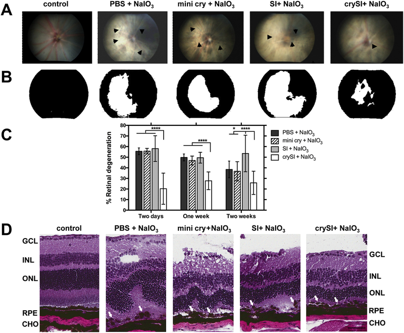
Fig. 2.
Intra-vitreal crySI protects against retinal degeneration caused by NaIO3 challenge in 129S6/SvEvTac mice. (A) Color fundus photography was used to compare therapeutic efficacy of crySI with controls in the mouse retina. Mice were pre-treated with 2 μL into the vitreous chamber with PBS, SI, crySI, or mini cry (250 μM) two days prior to intra-venous challenge with NaIO3 (33 mg/kg). Retinas were imaged one week later. Arrowheads indicate areas of retinal degeneration. (B) Fundus photographs were converted to greyscale and thresholded to identify the percentage area with significant degeneration (white) as a percent of the entire field (black). (C) NaIO3 challenge induced significant retinal degeneration in control mice pre-treated by two days, one week, and two weeks. For all pre-treatment periods, only crySI significantly reduced the area of retinal degeneration. Data are presented as mean ± SD (n = 6–10, *p < 0.05, ****p < 0.0001). (D) For animals pre-treated two days prior to NaIO3, H&E histopathology is presented to visualize retinal cryo-sections post-challenge. The epithelial monolayer was entirely disrupted and RPE cells showed a rounded, degenerative phenotype (see arrows) in all groups, except in the crySI pre-treated retinas. Predominant loss of RPE cells, distortion and thinning of the ONL and disorganization of the INL were also observed. Pre-treatment with crySI preserved retinal layers including RPE and photoreceptors (see arrows). GCL = Ganglion cell layer; INL = Inner nuclear layer; ONL = Outer nuclear layer; RPE = Retinal pigment epithelium; CHO = Choroid. Scale bar: 50 μm.

