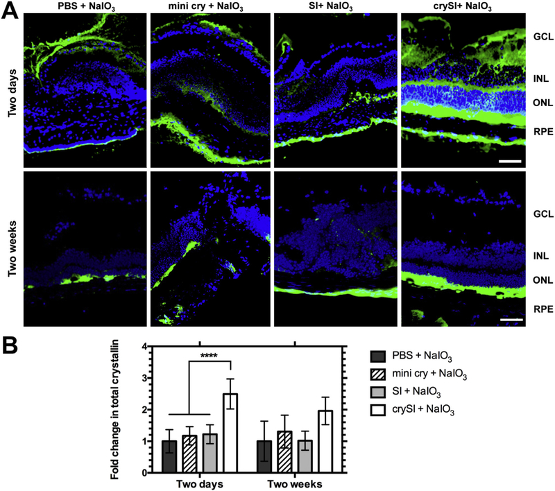
Fig. 6.
Intra-vitreal crySI enhances the accumulation of αB crystallin. 129S6/SvEvTac mice were pre-treated with PBS, mini cry, SI and crySI two days or two weeks prior to NaIO3 challenge. (A) Secondary immunofluorescence was used to detect both endogenous αB crystallin and residual exogenous crySI in cryo-sections of the retina. Due to antibody cross-reactivity, CrySI enhanced the staining for total αB crystallin detected in the RPE, INL, ONL layers in the two day pre-treatment groups (obtained 9 days after treatment with CrySI). In contrast, the two week pre-treatment retinas (obtained 6 weeks after treatment with CrySI) revealed a level and pattern of staining consistent with endogenous αB crystallin. Scale bar: 50 μm. (B) Quantification revealed that total crystallin level was significantly increased in the two-day pretreated crySI group compared to other controls. Data are shown as mean ± SD (n=5–7, ****p < 0.0001).

