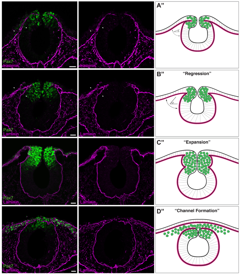
Figure 1. The basement membrane is remodeled during cranial neural crest EMT.
(A-D) Immunostaining for Pax7 (green) and laminin (magenta) in cross sections of HH8+ (A), HH9- (B), HH9 (C), and HH9+ (D) embryos. Arrowheads in (A’) and (B’) indicate apposed and non-apposed basement membranes, respectively. Scale bar, 20 μm. Schematics (A”-D”) summarize immunostaining data (n=12 sections, 3 embryos per stage), where green circles represent neural crest cells and magenta lines indicate the laminin-rich basement membrane. As development progresses from HH8+ to HH9-, a “regression” occurs, where the angle (θ) between the non-neural ectoderm basement membrane and the neuroepithelium basement membrane increases (A”-B”). As Pax7+ neural crest cells undergo EMT, a laminin channel forms by HH9+, creating a passage through which the cells are able to migrate.

