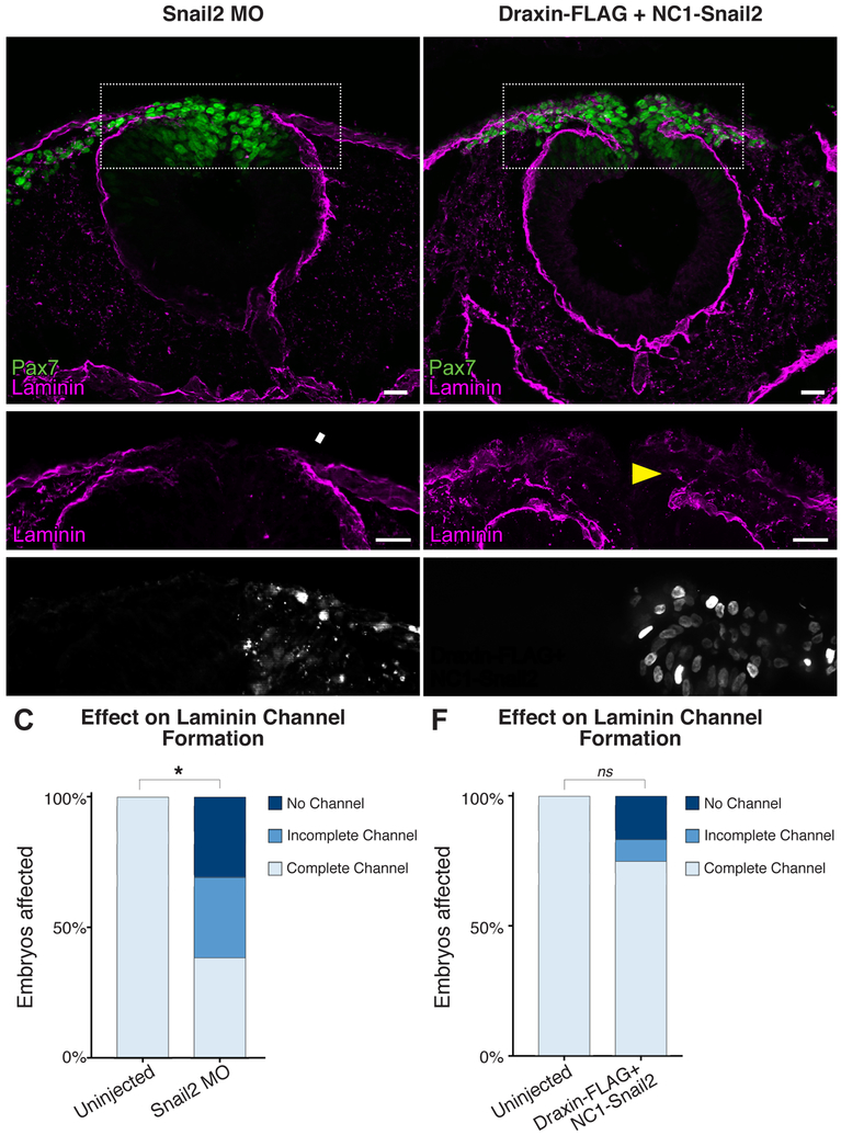
Figure 4. Modulating Snail2 alters laminin channel formation.
(A-B) Representative images for Pax7 (green) and laminin (magenta) immunostaining in embryos unilaterally electroporated with FITC-labeled translation-blocking Snail2 morpholino (MO). Scale bar, 20 μm. (C) Quantification of the effect of MO electroporation indicated Snail2 knockdown significantly reduced laminin channel formation. Box in (A) indicates enlarged area in (B), as indicated. White arrowhead highlights area of altered laminin distribution. *, p < 0.001, two-tailed t-test. ns, nonsignificant. (D-E) Representative images for Pax7 (green) and laminin (magenta) immunostaining in embryos unilaterally co-electroporated with NC1-Snail2 and Draxin-FLAG. Nuclear fluorescence in E’ indicates electroporated side. Scale bar, 20 μm. Box in (D) indicates enlarged area in (E), as indicated. Yellow arrowhead highlights rescue of channel formation. (F) Quantification of laminin channel formation indicated that NC1-Snail2 rescued the effect of Draxin-FLAG overexpression on channel formation. ns, nonsignificant (p = 0.082).

