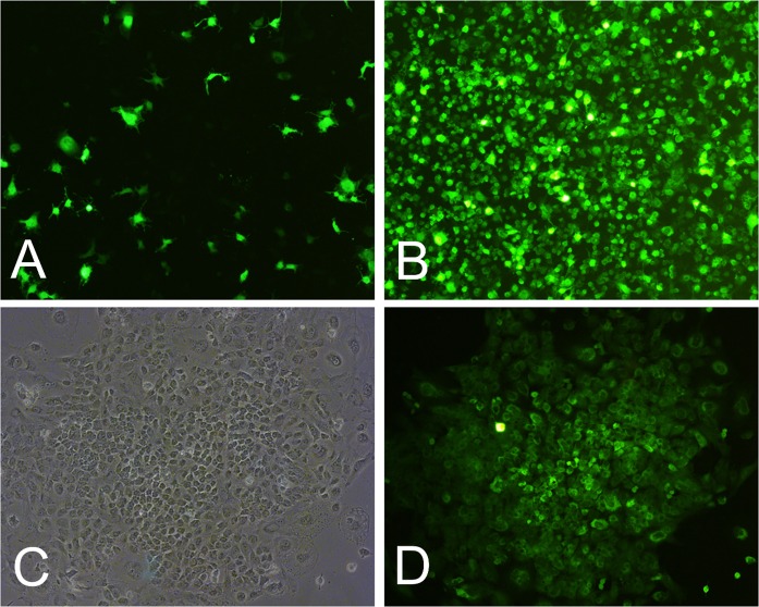
Fig. 1.
Intestinal epithelial cells infected with lentivirus and screened (× 100). a Cells observed by fluorescence microscopy 5 days post-infection. b Cells observed by fluorescence microscopy 15 days post-infection. c Monoclonal TIEC1s after screening. d Monoclonal cells observed by fluorescence microscopy

