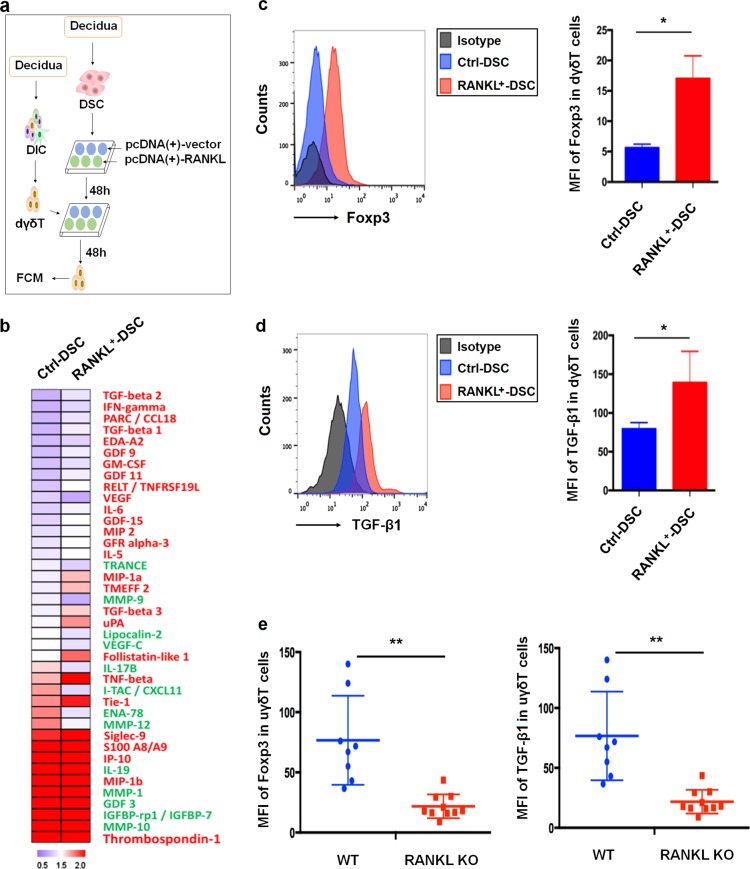
Fig. 4. RANKL from DSCs induce polarization of Foxp3 + regulatory γδT cells and TGF-β1 production.
a After co-culture with RANKL-overexpressed (RANKL+) or control (ctrl) DSCs at a 1:1 ratio for 48 h, the supernatants and decidual γδ T (dγδ T) cells were collected for protein array and flow cytometry analysis. b Heatmap of selected proteins differentially expressed in culture supernatants. c, d The median fluorescence intensity (MFI) of Foxp3 (c) and TGF-β1 (d) in dγδ T cells. e FCM analysis of MFI of Foxp3 and TGF-β1 in uterine γδ T cells from WT (n = 8) pregnant and RANKL−/− (n = 10). WT wild-type, KO knockout. Data are shown as the mean ± SD. *P < 0.05 and **P < 0.01 (Wilcoxon matched-pair signed-ranks test or Student’s t-test with correction)

