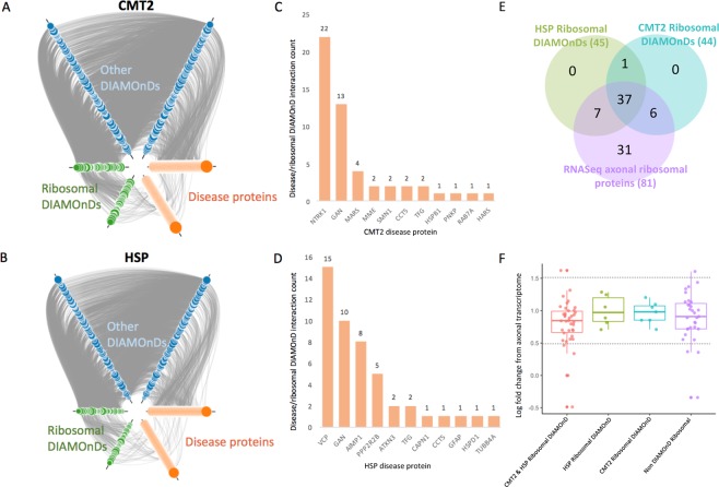
Figure 3.
Interactions within the expanded inherited axonopathy modules. (A,B) First degree interactions within CMT2 (A) and HSP (B) expanded modules. DIAMOnD proteins are separated into ribosomal proteins (green) and other (blue). Size of DIAMOnD nodes are proportional to DIAMOnD order of incorporation – smaller nodes were incorporated earlier. (C,D) First degree interaction counts between disease protein and ribosomal DIAMOnD proteins. (E) Relationships between HSP ribosomal DIAMOnD proteins, CMT ribosomal DIAMOnD proteins, and ribosomal proteins significantly localized to the axons of human motor neurons (q-value ≤ 0.1) (F). Distribution of log fold changes between axonal and soma differential localization.

