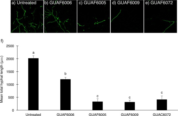
Figure 4.
Confocal laser scanning microscopy of FocuGFP-10 in bacterized soil. (a) Non-bacterized, (b) Chryseobacterium isolate GUAF6006, (c) Flavobacterium isolate GUAF6006, (d) Flavobacterium isolate GUAF6009, and (e) Flavobacterium isolate GUAC6072. Data are representative of nine images. (f) Mean total length of FocuGFP-10 hyphae in each camera field of view. Bars represent mean of three replications, and the different letters above the bars indicate statistically significant differences (P < 0.01, Tukey’s test).

