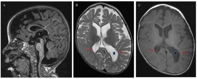
Figure 1.

Brain MRI patient 3 at 18 months of age: (A) Sagittal T1 MPRAGE –weighted image shows thinning of corpus callousm (white arrow). (B) and (C) axial T2 and T1-weighted images show brain volume loss, paucity white matter (red arrows) and asymmetry enlargement of lateral ventricles (star) left more than right. Skull deformity.
