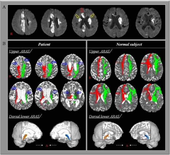
Fig. 1.

(A) Brain MR images at onset show multiple cerebral infarctions in both hemispheres including bilateral caudate nuclei (yellow arrows) and right cingulum (red arrow). (B) Results of diffusion tensor tractography for the ascending reticular activating system (ARAS). On 3-month diffusion tensor tractography, the neural connectivity in the upper ARAS between the thalamic intralaminar nucleus and the cerebral cortex is decreased in both prefrontal cortices and right cingulate cortex (blue arrows). By contrast, the lower ARAS between the pontine reticular formation and the thalamic intralaminar nucleus does not show significant abnormality compared with a normal subject (72 year-old female).
