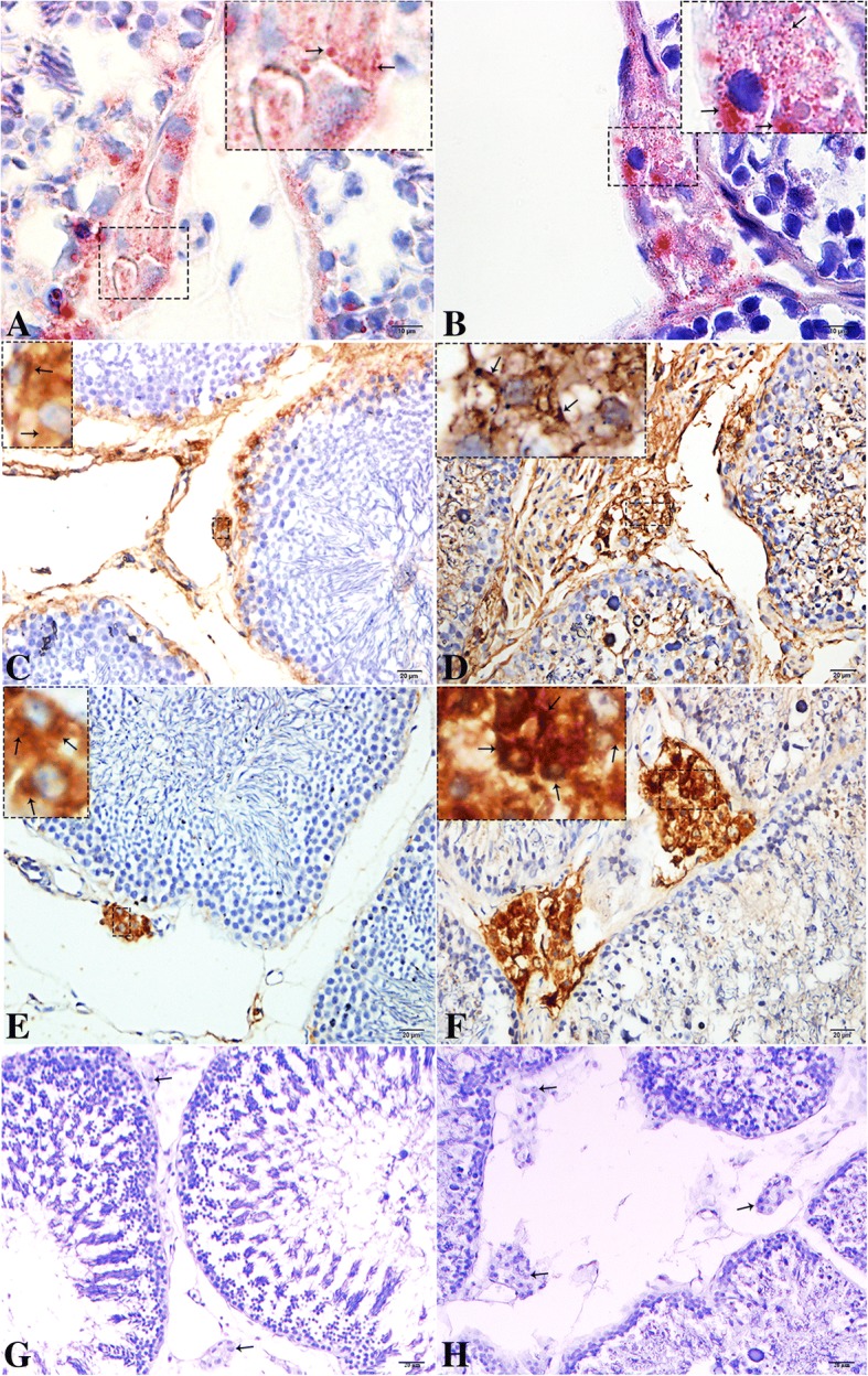
Fig. 2.

Light micrograph of Oil Red O staining and immunohistochemistry of vimentin, and 3β-HSD in the testis during resproductive and hibernation phase. (a-b) ORO staining of lipid droplets (arrow), (c-d) vimentin (e-f) 3β-HSD immunolocalization (arrow) and (G-H) negative control (arrow) in Leydig cell. A higher magnification is illustrated in the rectangular area. Scale bar = 10 μm (a-b) and 20 μm (c-h)s
