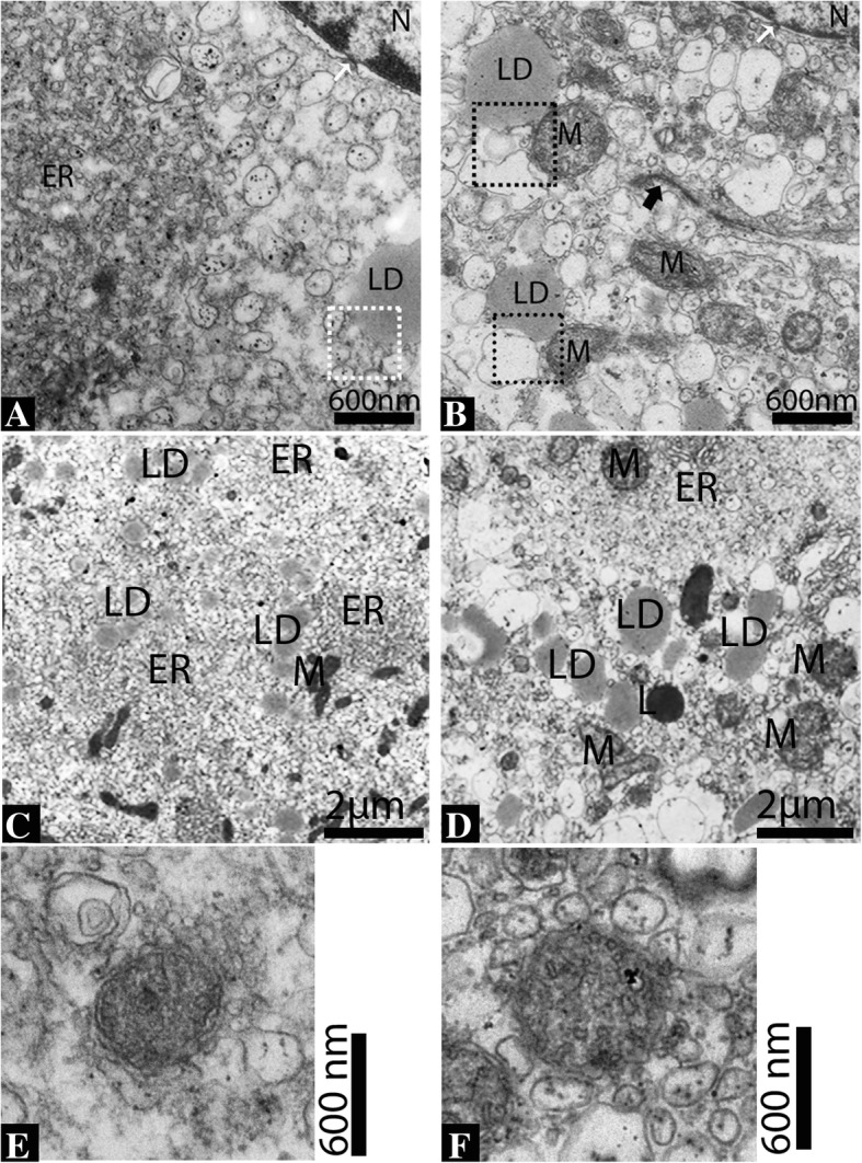
Fig. 6.

Ultrastructure of the Leydig cell. Abundant vesicular endoplasmic reticulum (a), and tubular endoplasmic reticulum and mitochondria (b) appear close to the nucleus and also show the nucleus pores (white arrow). Lipid droplets exhibit closely with vesicular endoplasmic reticulum during reproductive phase (c). In hibernation, the tubular endoplasmic reticulum, mitochondria and lysosome appeared to be in contact with the lipid droplets (d). The endoplasmic reticulum in contact with a lipid droplet (white dotted square) and with a lipid droplet and the mitochondria (black dotted square). Tight junctions between Leydig cell are also seen (black thick arrow). Mitochondria with well-developed inter cristae tubules (e-f). N: nucleus; ER: endoplasmic reticulum; LD: lipid droplets; M: mitochondria; L: Lysosome. Scale bar = 600 μm (a-b), 2 μm = (c-d) and 600 μm (e-f)
