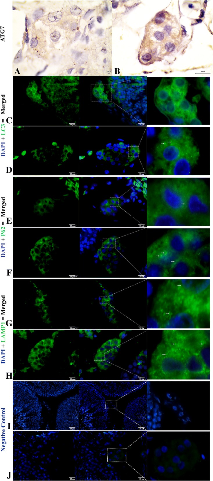
Fig. 7.

Immunofluorescent localization of autophagy markers in Leydig cells of the Chinese soft-shelled. Immunohistochemistry of ATG7 positive reactions in the Leydig cell (a-b). Immunofluorescence labeling of LC3, p62 and LAMP1 in the Leydig cell during reproductive (c, e and g) and during hibernation phase (d, f and h). Negative control; Reproductive (i) and hibernation phase (j). (White arrow): indicates positive localization. The higher magnification is illustrated by the rectangular area. Scale Bar = 10 μm (a-h)
