
Fig. 1.
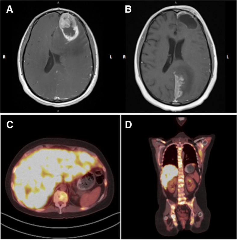
Axial MRI brain post administration of gadolinium a on initial presentation with a large contrast enhancing lesion and a necrotic core in the left frontal lobe with mass effect. b Images 2 weeks after completion of radiation therapy showing an enhancing lesion involving the paramedian left parietal-occipital lobe extending to the superior surface of the left cerebellar tentorium. c axial and d coronal FDG PET scan with hypermetabolic lytic bony lesions throughout the axial and proximal appendicular skeleton as well as innumerable hyperdensities in the liver concerning for metastatic disease