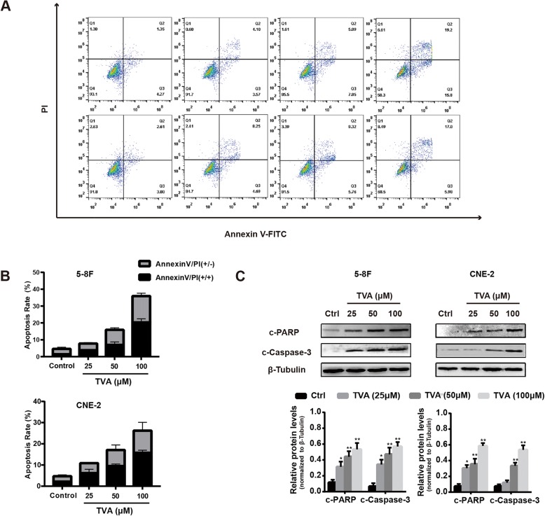
Fig. 2.
TVA treatment induces apoptosis in NPC cells. a. Apoptosis was analyzed by flow cytometry with PI and annexin V-FITC staining after 5-8F and CNE-2 cells were treated with TVA for 24 h at the indicated concentrations. b. The percentage of apoptotic cells was calculated as the apoptosis rate. c. Cleaved PARP (c-PARP) and cleaved caspase-3 (c-Caspase-3) protein levels after TVA treatment for 24 h. β-tubulin was used as an internal control. *P<0.05 and **P<0.01 versus the control group

