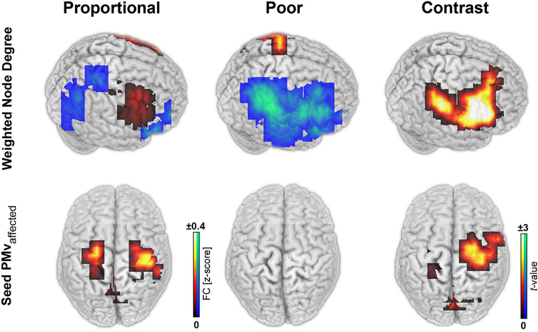
Figure 4. EEG network correlates of proportional recovery.
The weighted node degree (WND) of resting-state functional connectivity was disrupted in the affected hemisphere of patients with POOR (blue). Conversely, patients with PROP showed large WND (red-yellow) (A). Enhanced functional connectivity was observed in particular between the ventral premotor (PMv) and primary motor area of the affected hemisphere of patients with PROP (B). All stroke lesions are aligned to the right hemisphere (shown right).

