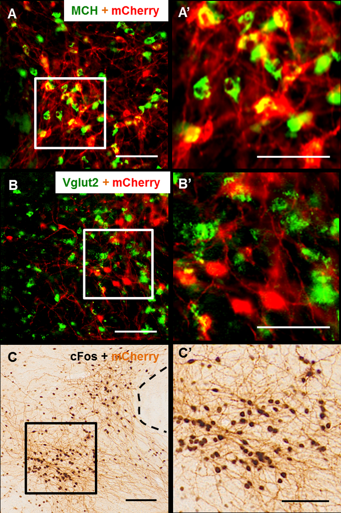
Figure 2: Chemoactivation of MCH neurons by CNO in MCH-Cre and MCH-Vglut2KO mice.

(A, B) Representative brain sections, labelled for mCherry (red) and MCH (green in A, A’) or Vglut2 mRNA (green in B, B’) from a MCH-Vglut2KO mouse injected with AAV-hM3Dq into the LH. AAV injections resulted in specific expression of hM3Dq in MCH neurons (>90% of hM3Dq-mcherry expressing neurons were positive for MCH). These mCherry-ir neurons did not contain Vglut2 mRNA further indicating that hM3Dq was expressed in the MCH neurons lacking Vglut2. (C, C’) Representative brain section from a MCH-Vglut2KO mouse labelled for cFos (black) and mCherry (brown) using DAB immunohistochemistry. This mouse was intraperitoneally injected with CNO (0.3 mg/kg) and sacrificed after 3 h. CNO induced cFos in almost all the mCherry+ neurons indicating their activation. Squares in A, B and C indicate the region magnified in the A’, B’ and C’ respectively. Scale bars - 100 µm.
