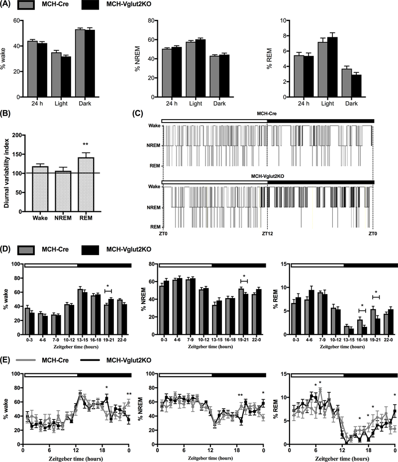
Figure 3: Specific elimination of glutamate neurotransmission from MCH neurons alters diurnal variation of REM sleep.

(A) Percentages of wake, non-rapid eye movement sleep (NREMs) and rapid eye movement sleep (REMs) in MCH Cre (n=10; gray bars) and MCH-Vglut2KO mice (n=12; black bars) during the entire 24-h or specifically during the light and dark periods. (B) Diurnal variability index, a measure of diurnal amplitude of wake, NREMs and REMs in MCH-Vglut2KO mice normalized to control data (100%) from MCH-Cre mice (horizontal line) (Mann-Whitney U test; **p < 0.001. (C) Representative 24-h hypnograms from one MCH-Cre (controls, upper trace) and one MCH-Vglut2KO mouse (lower trace). MCH-Vglut2KO mice displayed significant REMs suppression during the dark period compared to MCH-Cre mice. (D) and (E) Percentages of wake, NREMs and REMs in 3-h bins (D) and 1-h bins (E) in MCH-Cre (gray lines or bars) and MCH-Vglut2KO mice (black lines or bars) *p < 0.05; **p < 0.01; Multiple t-test with Holm-Sidak method. In C, D and E, white horizontal bar indicates the light period (ZT0-12) and the black horizontal bar depicts the dark period (ZT12-0). All data are mean ± SEM.
