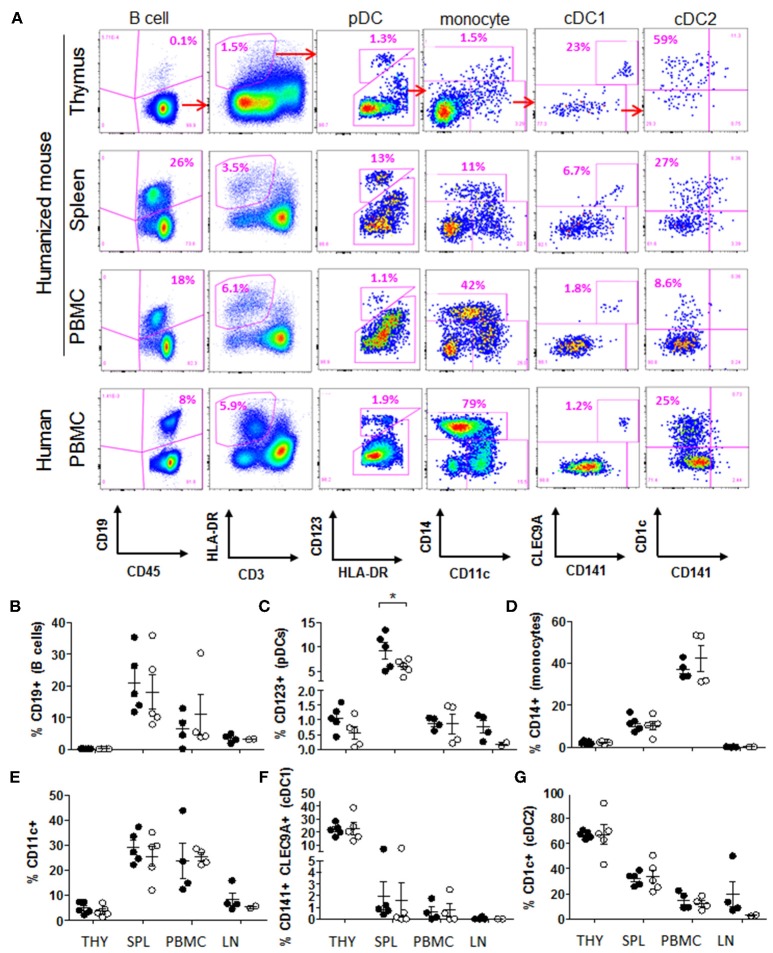
Figure 3.
HSCs generate all major human APC populations in humanized mice. (A) Flow cytometric identification of major type of APCs in the thymic graft, spleen and peripheral blood of humanized mice at end point (example from Peptide group); and in the blood of a healthy human donor. (B–G) Relative frequency of APC populations in Peptide group (closed circles) and Control group (open circles): (B) CD19+ B cells (gated on CD45+ cells), (C) CD123+ pDCs (gated on CD45+ HLA-DR+ CD3− cells), (D) CD14+ monocytes (gated on CD45+ HLA-DR+ CD123− cells), (E) CD11c+ cells (gated on CD45+ HLA-DR+ CD123− cells), (F) CD141+ CLEC9A+ cDC1 cells (gated on CD11c+ cells), and (G) CD1c+ cDC2 cells (gated on CD11c+ CLEC9A− cells). All graphs show the mean ± SEM from n = 5 mice per group (thymus and spleen); n = 4 mice per group (PBMC); n = 4 (lymph nodes from Peptide group) and n = 2 (lymph nodes from Control group). t-test comparison showed no difference between groups, except where indicated. THY, thymus; SPL, spleen; PBMC, peripheral blood mononuclear cells; LN, lymph nodes.

