Figure 3.

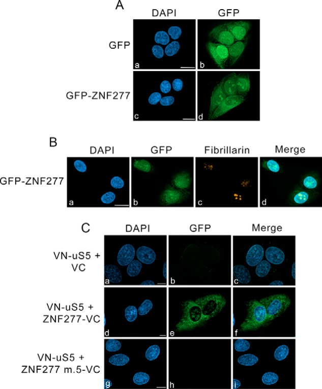
ZNF277–uS5 complexes localize to the cytoplasm and nucleolus. A, U-2 OS cells induced to expressed GFP (panels a and b) and GFP–ZNF277 (panels c and d) were fixed and analyzed by direct fluorescence. DNA staining with 4′,6-diamidino-2-phenylindole (DAPI) shows the nucleus of each cell (panels a and c). Bar, 10 μm. B, U-2 OS cells induced to express GFP–ZNF277 were simultaneously analyzed by direct GFP fluorescence (panel b) and immunostaining for the nucleolar marker fibrillarin (panel c). DNA staining with DAPI shows the nucleus of each cell (panel a). Bar, 10 μm. C, representative BiFC images showing interaction between VN-uS5 and ZNF277-VC in living human cells. U-2 OS cells that coexpressed VN-uS5 with either the VC control (panels a–c), WT ZNF277-VC (panels d–f), and ZF#5 mutant ZNF277-VC (panels g–i) were fixed and analyzed by direct fluorescence. DNA staining with DAPI shows the nucleus of each cell (panels a, d, and g). Bar, 10 μm.
