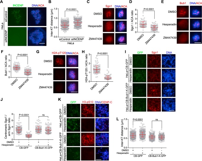
Figure 1.
Inhibiting Aurora B kinase activity causes centromeric cohesion defects, which can be rescued by centromere-tethered Bub1. A and B, HeLa cells transfected with control or INCENP siRNA were treated with nocodazole for 3 h. Mitotic cells collected by shake-off were spun on slides and stained with INCENP antibodies, ACA, and DAPI (A). The inter-KT distance was measured on over 886 chromosomes in 20 cells (B). C–H, HeLa cells were treated for 1 h with nocodazole and MG132 together with DMSO or the indicated Aurora B inhibitors. Cells were then stained with DAPI, ACA, and antibodies for Sgo1 (C), Bub1 (E), and H2A-pT120 (G). The immunofluorescence intensity ratios of centromeric Sgo1/ACA (D), Bub1/ACA (F), and H2A-pT120/ACA (H) were determined on 90–123 chromosomes in 20 cells. I–L, HeLa cells stably expressing CB-GFP or CB-Bub1-K-GFP were treated for 1 h with nocodazole and MG132 together with DMSO or Hesperadin. Mitotic chromosome spreads were stained with DAPI and antibodies for Sgo1, H3-pS10, and CENP-C. Example images are shown (I and K). The immunofluorescence intensity ratio of centromeric Sgo1/arm Sgo1 was determined on at least 124 chromosomes in 20 cells (J). The inter-KT distance was measured on over 759 chromosomes in 20 cells (L). Means and error bars representing S.D. are shown (unpaired t test). Scale bars, 10 μm. See also Fig. S1. ns, not significant.

