Figure 7.
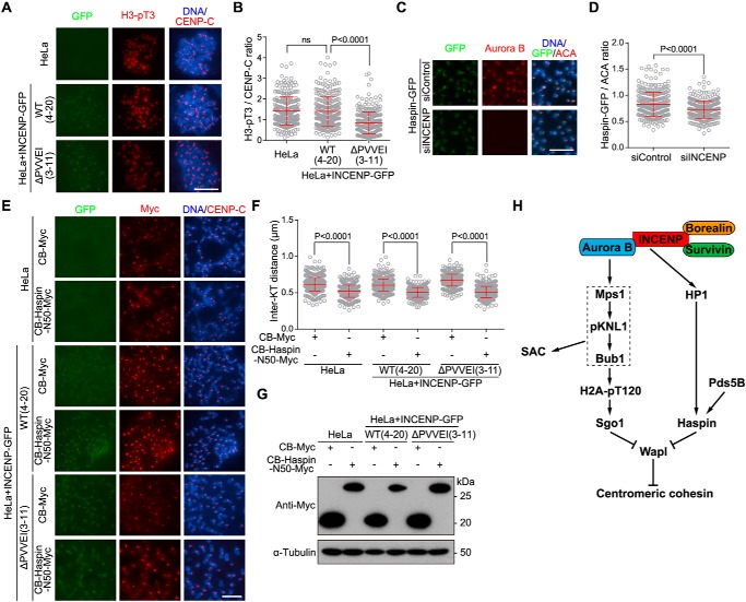
The INCENP–HP1 interaction promotes centromeric localization of Haspin to antagonize Wapl. A and B, HeLa cells and the indicated stable cell lines were treated with nocodazole for 3 h. Mitotic cells were spun on slides and stained with DAPI and antibodies for H3-pT3 and CENP-C (A). The centromeric H3-pT3/CENP-C immunofluorescence intensity ratio was determined on 273–285 chromosomes in 20 cells (B). C and D, HeLa cells stably expressing Haspin-GFP were transfected with control or INCENP siRNA and treated with nocodazole for 3 h. Mitotic chromosome spreads were stained with DAPI and antibodies for GFP, Aurora B, and ACA (C). The centromeric GFP/CENP-C immunofluorescence intensity ratio was determined on around 320 chromosomes in 30 cells (D). E–G, HeLa cells and the indicated stable cell lines were transfected with CB-Myc or CB-Haspin-N50-Myc and treated with nocodazole for 3 h. Mitotic chromosome spreads were stained with DAPI and antibodies for Myc and CENP-C (E). The inter-KT distance was measured on 560–640 chromosomes in 25 cells (F). The asynchronous cell lysates were immunoblotted (G). H, model for the Aurora B kinase activity–dependent and –independent roles for the CPC in protecting sister chromatid cohesion at mitotic centromeres. Aurora B kinase activity–dependent localization of the Mps1–pKNL1–Bub1 signaling cascade to the kinetochore promotes centromeric cohesion through phosphorylating H2A and recruiting Sgo1 to centromeres. Aurora B kinase activity–independent INCENP–HP1 interaction protects centromeric cohesion through promoting the centromeric localization of Haspin. Both Sgo1 and Haspin antagonize Wapl activity in releasing cohesin from mitotic centromeres. Means and error bars representing S.D. are shown (unpaired t test). Scale bars, 10 μm. ns, not significant.

