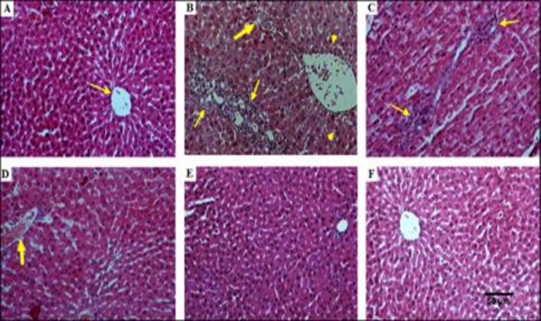
Figure 3.
A: Histopathological study of liver tissue and effect of hydroalcholic extract of Zataria. multiflura in liver tissues in diabetic rats. H&E staining with magnification at X100
A: Healthy control rats with normal hepatocytes and normal central vein (thin arrow). B: Diabetic control rats with an increment in infiltration of the lymphocytes (thin arrows), congested and edematous portal vein and hemorrhage (thick arrow). Also, vacuolation in the cytoplasm of the hepatocytes appeared as indistinct clear vacuoles (pick arrows). C: Diabetic rats treated with Z. multiflora 250mg/kg showed lymphocytic inflammation and vacuolization of cytoplasm (thin arrow). D: Diabetic rats treated with Z. multiflora 500 mg/kg showed fewer pathological changes and improved liver architecture. E: Diabetic rats treated with Z. multiflora 1000 mg/kg with relative restoration of the liver architecture and normal hepatocytes. F: Non-diabetic rats treated with Z. multiflora 1000 mg/kg with normal liver histopathological features.

