
Figure 1.
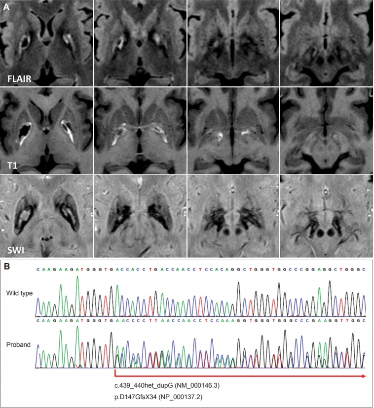
Brain MR images and the DNA sequence of the neuroferritinopathy patient. A: Fluid-attenuated inversion recovery (FLAIR) MR images show a trilamellar cystic lesion in the pallidum. T1-weighted and susceptibility-weighted (SWI) MR images show a cystic core and a surrounding rim of iron deposition. B: Sanger sequencing results of the proband showing a novel frameshift mutation (c.439_440het_dupG, NM_000146.3) in the FTL1 gene.