Figure 5.
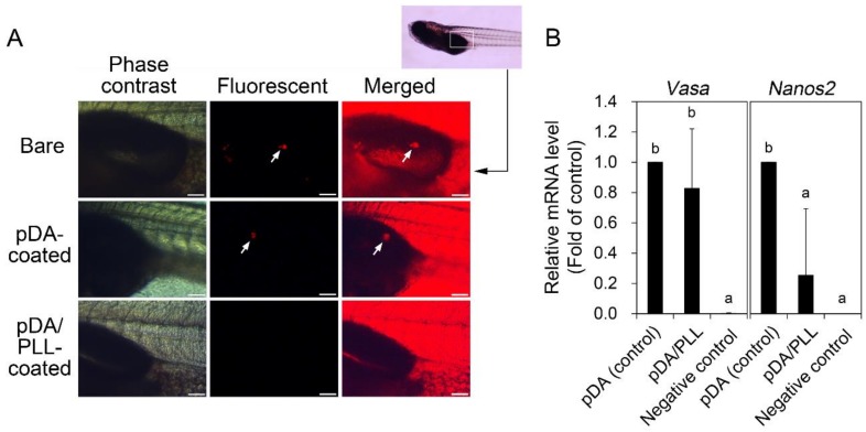
Effects of pDA or pDA/PLL coating on gonadal colonization and relative mRNA expression of vasa and nanos2 genes of ovarian germline stem cells (OGSCs) after culture. (A) Gonadal colonization of OGSCs cultured on pDA- and pDA/PLL-coated dishes. The OGSCs cultured for 10 days on pDA- and pDA/PLL-coated dishes were labeled with fluorescent dye (PKH26) and intraperitoneally transplanted into 11 days-post-fertilization (dpf) larvae. After 9 days, gonadal colonization of the transplanted OGSCs was observed in the larvae transplanted with the OGSCs cultured on non-treated and pDA-coated dishes. In contrast, no colonization was observed in larvae transplanted with the OGSCs cultured on pDA/PLL-coated dishes. Arrows indicate the OGSCs colonized. Scale bar = 100 μm. (B) Relative mRNA expression of vasa and nanos2 genes between the enriched OGSCs cultured on pDA and pDA/PLL-coated dishes. The enriched OGSCs were cultured on pDA- or pDA/PLL-coated dishes and then the cells were subjected to quantitative reverse transcription polymerase chain reaction (qRT-PCR) analysis. A significant decrease of nanos2 expression was observed in the cells cultured on pDA/PLL-coated dishes compared to the cells cultured on pDA-coated dishes, whereas no significant difference was detected in vasa expression between two cell populations. ab Different letters indicate significant difference, p < 0.05. An Oryzias latipes embryonic cell line was used as a negative control.

