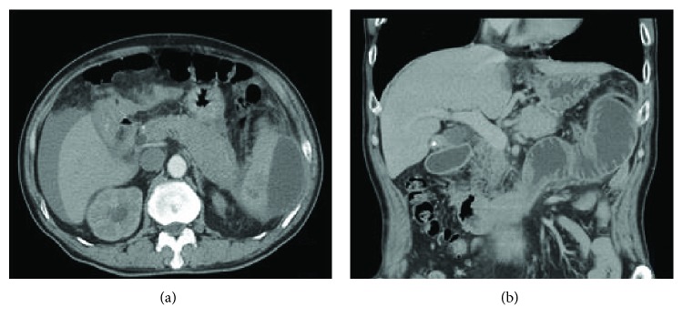
Figure 2.
CT scan of patient no. 4. (a) Axial section of the CT scan showed a hematoma in the lower pole and lateral spleen area combined with the enlargement of the body and tail of the pancreas and free fluid around the liver and spleen. (b) Coronal section of the CT scan showed an obstruction of the small intestine.

