. 2019 Feb 7;114:e180432. doi: 10.1590/0074-02760180432

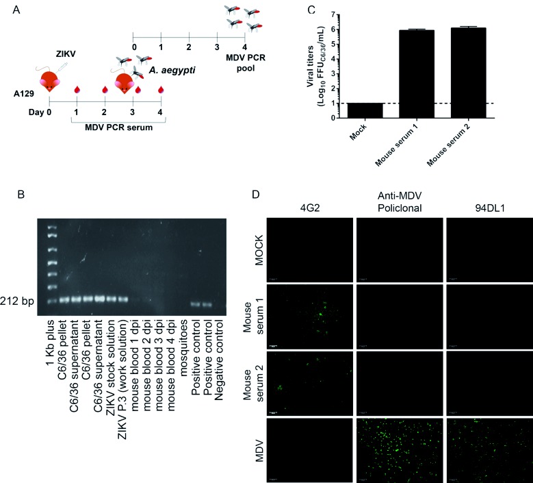
Fig. 3: infection of A129 mice provide a reliable strategy for clearance of mosquito densovirus (MDV)-contamination. (A) Experimental design for in vivo assays. (B) Agarose gel showing reverse transcription-PCR (RT-PCR) amplification of a 212 bp fragment from MDV in the blood of A129 mice infected with 4x10⁶ PFU of Zika virus (ZIKV) strain A at different days post infection (dpi) and in Aedes aegypti mosquitoes that fed on infected animals. As a control, ZIKV strain A stocks and C6/36 cells (pellets and culture supernatants were used) together with a plasmid control were tested for MDV. (C) Viral titers in mice sera three days post-infection. The sera from two mice were tested in biological replica. (D) Immunofluorescence assay of C6/36 cells infected with ZIKV strain A recovered from mice sera three days post-infection. C6/36 cells were infected with mice sera at a multiplicity of infection (MOI) of 1 and after three days stained with 4G2 monoclonal antibody, anti-MDV mouse polyclonal serum, and anti-MDV monoclonal antibody (clone 94DL1). As a positive control, C6/36 cells were infected with MDV BR/07 at a multiplicity of genome (MOG) of 0.01 for 72 h.

Fig. 3: