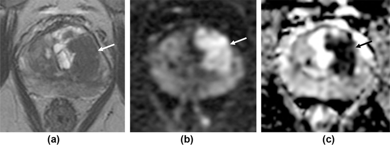
Figure 2.

Differences inT2 scoring for high probability targets. A 77-year-old patient with a PSA of 17 ng/ml. (a)T2W1 shows a large (2.5 × 1.5 cm) lesion centred in the left TZ at the level of the mid-gland (arrow), with matching restricted diffusion (b-c). P1-RADS v1 scores: 4 for T2 as no features of ECE and no broad capsular contact, 5 for DW1. P1-RADS v2 overall score 5: T2 is the dominant sequence and the lesion is >1.5 cm, despite no features of ECE. Subsequent targeted biopsy confirms Gleason 4+5 disease (90% core involvement) in the left medial mid-gland and Gleason 5+5 (60% involvement) in the left posterior lateral gland.
