
Figure 4.
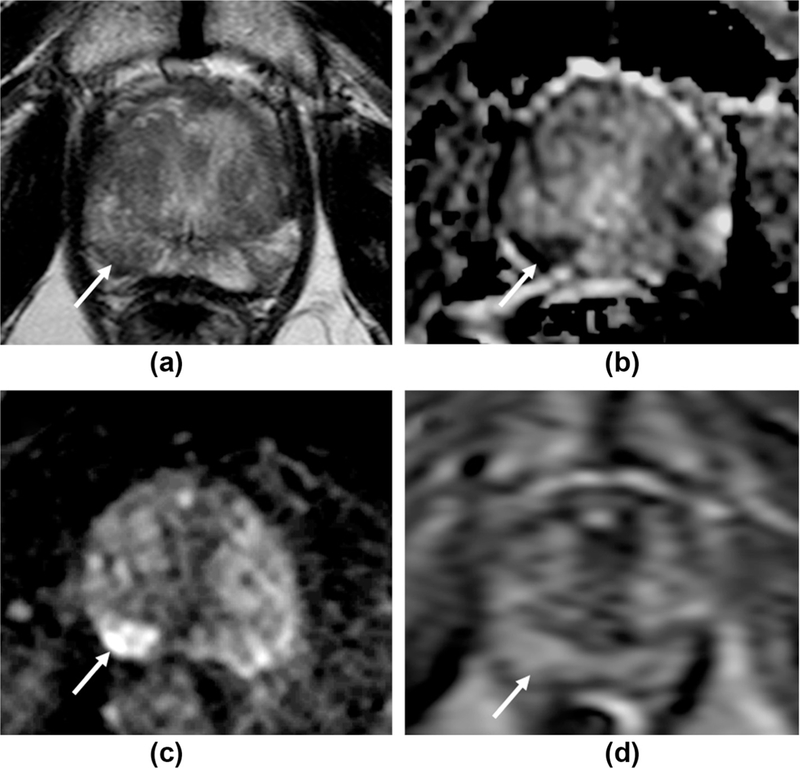
False-negative DCE, correctly evaluated as likely tumour using PI-RADS v2 criteria. 60 year-old patient with a PSA of 4.48 ng/ml. a: T2W1 imaging shows a focal area of intermediate/low signal the right mid/apex PZ (arrows), with marked matching restricted diffusion (b, c). d: The region shows diffuse, but no focal or early enhancement on DCE-MRI (arrow), with a Type I curve. PI-RADS v1 scores: 3 for T2, 5 for DWI, 1 for DCE, summed score=9. PI-RADS v2 overall score=5 (DWI is the dominant PZ sequence) despite being “negative” with DCE. Targeted biopsy demonstrated a Gleason 3+3 tumour in 10% of the cores.