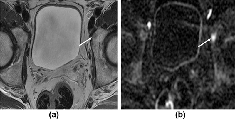
Figure 5.

High b-value images aid detection of bone metastases. A 68-year-old man with rising PSA, 2 years post-prostatectomy. (a) Axial T2W1 shows a subtle low intensity area in the left acetabulum (arrow). (b) Axial b=1400 DWI sequence; the lesion demonstrates restricted diffusion and appears more conspicuous on these high b-value images.
