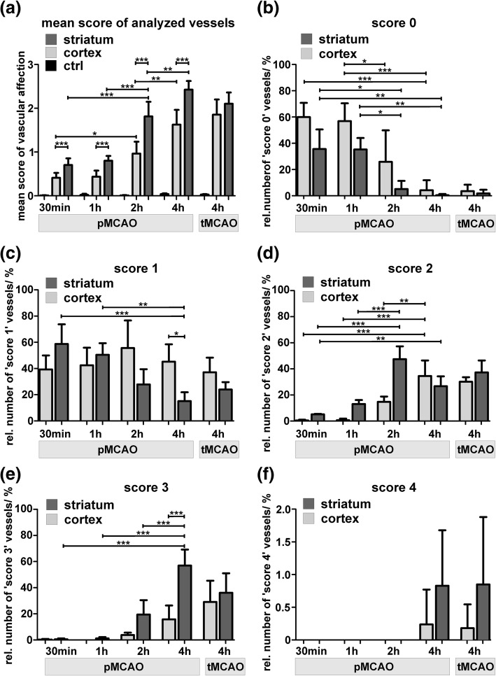
Fig. 4.
At the level of electron microscopy, the described scores of vascular damage are used to quantitatively address vascular alterations at an ultrastructural level. a Mean score of analyzed vessels in contralateral control areas (ctrl), and ischemia-affected striatal and cortical areas of 30 min, 1 h, 2 h and 4 h pMCAO animals. Further, analysis included 4 h tMCAO animals, representing the reperfusion scenario. b-f Comparison of the relative numbers of ischemia-affected vascular damage (score 0–4). Importantly, the relative number of vessels showing an unaffected endothelial cells (score 0) is found to decrease from 30 min to 4 h pMCAO animals. Of note, as soon as 30 min after ischemia onset, up to 60% of the analyzed vessels show signs of an endothelial edema (c, score 1). In line, more severe scores (score 2 & 3) are found to be significantly increased when comparing 30 min, 1 h, 2 h and 4 h pMCAO animals. f An extravasation of erythrocytes was restricted to 4 h pMCAO and tMCAO animals, but appeared to be a rare event. Of note, for all the described scores, a direct comparison between 4 h pMCAO and 4 h tMCAO animals did not reveal statistically significant differences. * p < 0.05, ** p < 0.01, *** p < 0.001; 30 min, 1 h, 2 h pMCAO and 4 h tMCAO: n = 4; 4 h pMCAO: n = 5; ANOVA followed by Bonferroni’s multiple comparison test. Data are given as means. Error bars indicate SD

