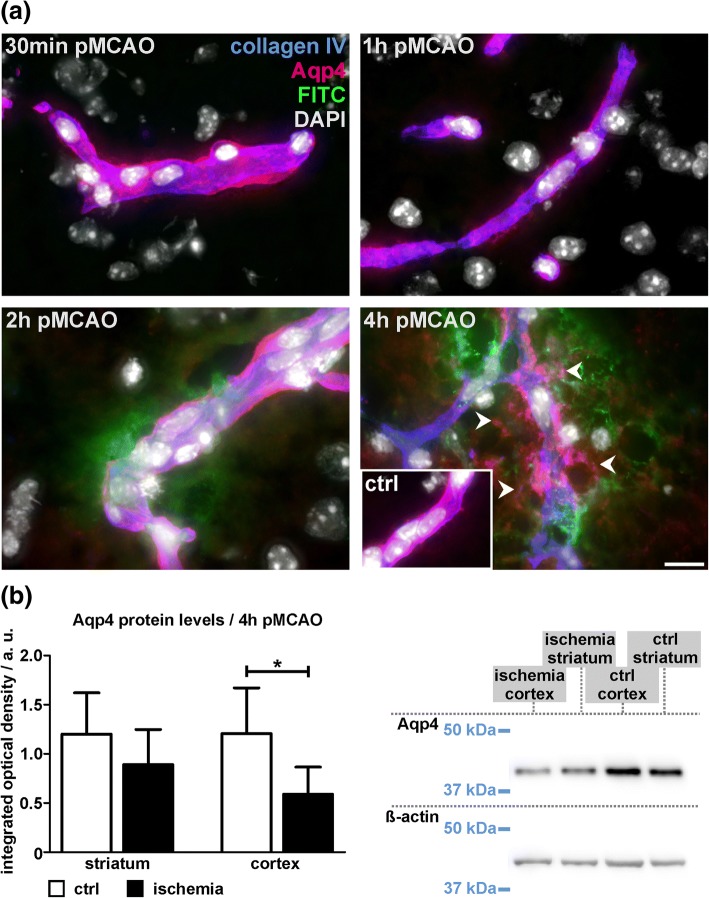
Fig. 7.
a Representative micrographs showing immunofluorescence labeling of vascular basement membranes (collagen IV) and aquaporin 4 (Aqp4) to illustrate the ischemia-associated affections of juxtavascular astrocytes. Again, an extravasation of FITC-albumin (FITC) is observed in 2 h and 4 h pMCAO animals. In contralateral control regions (ctrl, inset) Aqp4 expression is highly polarized and confined to astrocytic endfeet directly adjacent to the vascular basement membrane. Importantly, this polarization is lost around vessels showing FITC-albumin extravasation in 4 h pMCAO animals. Although FITC-albumin extravasations are also observed in 2 h pMCAO animals, an astrocytic depolarization is not observed, matching the observations from 1 h and 30 min pMCAO animals. Nuclei are visualized with DAPI. Scale bar: 10 μm. b Western Blot analysis reveals significantly decreased Aqp4 protein levels in cortical areas of 4 h pMCAO animals (p = 0.026), while this difference failed to reach statistical significance in the striatum. n = 6, Student’s t-test. Data are given as means. Error bars indicate SD

