. 2019 Jan 7;19(3):503–512. doi: 10.1039/c8lc01259h

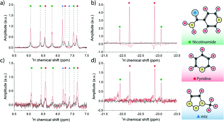
Fig. 3. (a and b) Single acquisition 1H spectra of the SABRE mixture (25 mM pyridine, 25 mM nicotinamide, 26 mM mtz, 2.4 mM Ir-IMes catalyst) performed at B = 11.7 T and T = 30 °C. The SABRE measurement (in red) was recorded immediately after thoroughly bubbling the sample solution with a 90% p-H2 enriched mixture for 20 s. The reference measurement at thermal equilibrium is plotted in black. The analyte signal peaks are shown on the left in (a), and the di-hydritic region to the right in (b). (c and d) 16-fold averaged 1H spectra for the high-field SABRE experiment performed at B = 11.7 T and T = 30 °C and under active microflow conditions. The flow rate was set at the optimal value of 60 μL min–1. The reference measurement, in black, was recorded at rest. Peak assignment follows the colour scheme reported on the right edge of the figure, i.e. nicotinamide peaks are labelled with green dots, pyridine with red, and mtz with blue in plots (a) through (d).

Fig. 3