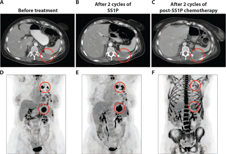
Fig. 5. Tumor response to post-SS1P chemotherapy in a patient with pleural mesothelioma.
(A to C) Representative axial CT images of the chest for patient 9 before treatment (A), after two cycles of SS1P (B), and after two cycles of post-SS1P chemotherapy (C). Areas of tumor involvement are indicated by red circles. (D to F) [18F]FDG PET images from time points corresponding to (A) to (C), respectively. Areas of tumor involvement are indicated by red circles. (F) Image showing a marked reduction of [18F]FDG uptake in the highlighted areas after post-SS1P chemotherapy, compared to after two cycles of SS1P (E). Intense bone marrow metabolic activity in the axial and appendicular skeleton in (F) is secondary to recent administration of granulocyte colony-stimulating factor to prevent chemotherapy-induced neutropenia.

