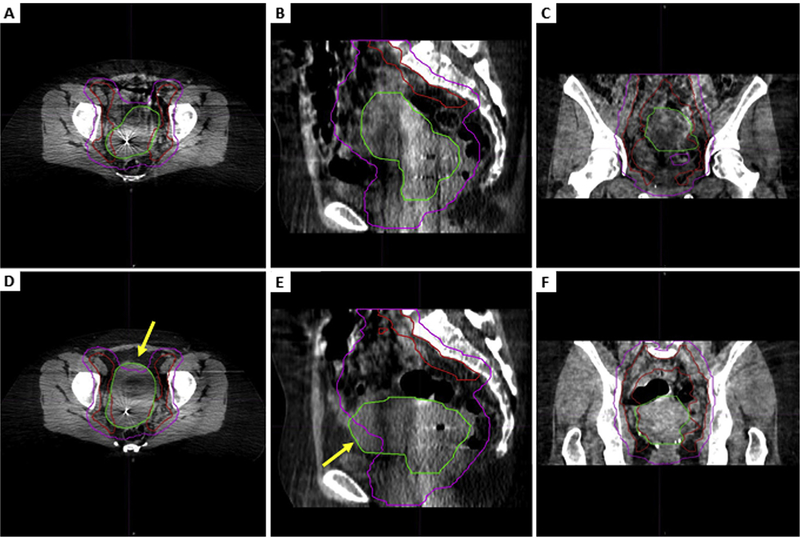
Fig. 1.
Representative cone beam computed tomography slices showing a fully encompassed clinical target volume (CTV) compared with a partially missed CTV. Axial (A), sagittal (B), and coronal (C) views of CTV comprising gross tumor, cervix, and uterus (CTV1) (green) and CTV comprising pelvic lymph nodes (CTV3) (red) that are completely encompassed within the 95% isodose structure (pink) and axial (D), sagittal (E), and coronal (F) views from a scan with an anterior CTV1 miss in the uterine body. The arrows point to the missed CTV1. (A color version of this figure is available at www.redjournal.org.)

