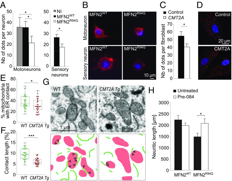
Fig. 3.
MFN2R94Q affects neuronal ER–mitochondria contacts in vitro and in vivo. (A) Quantification of ER–mitochondria contacts using PLA at 6 dpi in motor (n = 5 independent cultures) and sensory neurons (n = 3), infected with AAV6-hsyn-hMFN2WT or AAV6-hsyn-hMFN2R94Q (n = 6 independent cultures). Data are expressed as the mean number of contacts ± SEM per neuron detected by a PLA. Statistical analysis: repeated measures one-way ANOVA with Tukey’s post hoc test (motoneurons) and two-tailed paired Student’s t test (sensory neurons). NI, noninfected motoneurons. (B) Photomicrographs of PLA between the ER protein IP3R and the mitochondrial protein VDAC1 in motor and sensory neurons. Red dots indicate ER–mitochondria proximity. Nuclei are stained with DAPI. (C) Quantification of ER–mitochondria contacts using PLA in fibroblasts from control and CMT2A-R94Q individuals (n = 39 and n = 40 cells, respectively, from two independent cell lines for each condition). Data are expressed as the mean number of contacts ± SEM per cell. Statistical analysis: two-tailed unpaired Student’s t test. (D) Photomicrographs of control and CMT2A human fibroblasts. Intracellular red dots indicate the presence of ER–mitochondria proximity revealed by the PLA. Nuclei are stained with DAPI. (E) Electron microscopy quantification of the percentage of mitochondria with ER contact in motoneuron soma in the lumbar spinal cord of 12-mo-old WT (n = 34 motoneurons from three mice) and CMT2A Tg mice (n = 33 motoneurons from three mice). Statistical analysis: two-tailed unpaired Student’s t test. (F) Analysis of the length of the mitochondria–ER contacts, expressed as the percentage of the mitochondrial perimeter (n = 34 motoneurons from three WT mice and n = 33 motoneurons from three CMT2A Tg mice). Statistical analysis: two-tailed unpaired Student’s t test. (G) Electron microscopy pictures illustrating ER–mitochondria contacts in motoneuron soma of WT and CMT2A Tg mice. ER segments are delineated in green and mitochondria are shown in red. Arrowheads indicate the mitochondria–ER contacts. (H) Neuritic length quantified at 6 dpi in motoneurons overexpressing either MFN2WT or MFN2R94Q. Motoneurons treated with a SIGMAR1 agonist (Pre-084, 50 nM) are compared with the untreated condition. Note the significant rescue of neuritic length in MFN2R94Q motoneurons treated with Pre-084. Mean values are obtained from four independent cultures. Statistical analysis: two-way ANOVA (group × treatment) with Sidak post hoc test. *P < 0.05, ***P < 0.001.

