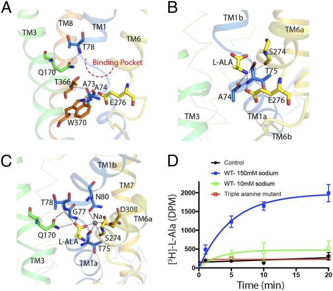
Fig. 3.
Gating interactions and substrate binding site in AgcS. (A) A close-up view of gating interaction network around the binding pocket in AgcS with l-alanine bound. (B) Hydrogen bonds between amino group of the substrate and the residues of AgcS. (C) Hydrogen bonds and ionic interactions in the hydrophilic part of binding pocket are depicted as dashed lines. Bound Na+ ion shown as a gray sphere. (D) The uptake ability for missense mutant (triple alanine mutation at N80A, S274A, and D308A) compared with that of the WT protein.

