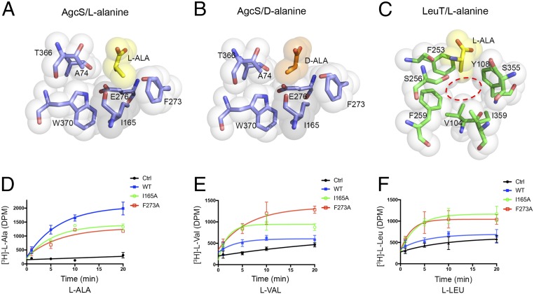
Fig. 4.
Substrate selectivity in AgcS is dictated by the size of the binding pocket. Comparison of the hydrophobic pocket of alanine-bound AgcS and LeuT (Protein Data Bank [PDB]: 2qei). (A–C) Van der Waals surfaces for alanine substrate and interacting residues are shown as colored spheres. The binding pocket of AgcS (dashed ring in C) is very small and only glycine or alanine can fit while in LeuT, where substrate selectivity is broader, the site is much larger to accommodate larger amino acids. (D–F) AgcS mutants I165A and F273A lose substrate selectivity allowing valine and leucine to be transported. Nonspecific uptake was assessed by using protein-free liposomes under identical conditions as described in Methods Summary. Ctrl, control.

