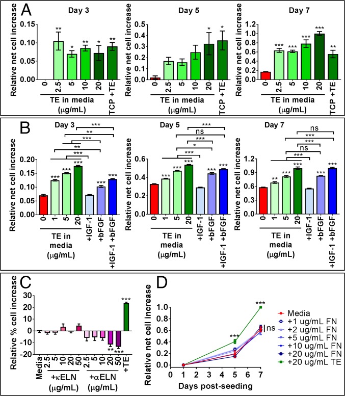
Fig. 3.
MSC proliferation in media with tropoelastin in solution. (A) Cells were grown on TCP in media supplemented with increasing concentrations of soluble tropoelastin or on tropoelastin-coated TCP in normal media. Panels show relative net cell increase at 3, 5, and 7 d postseeding. Asterisks above individual columns depict statistical differences from the no-tropoelastin control. (B) Cells were cultured on TCP in normal media or in media supplemented with tropoelastin or growth factor(s). Panels show relative net cell increase at 3, 5, and 7 d postseeding. Asterisks directly above the data columns indicate statistical differences from the normal media control. (C) Cell proliferation for 7 d in normal media or in media supplemented with κELN, αELN, or tropoelastin. Asterisks indicate statistical differences from the normal media control. (D) Cells were grown for up to 7 d in normal media or media supplemented with fibronectin or tropoelastin in solution. Asterisks denote statistical differences from the normal media control. *P < 0.05; **P < 0.01; ***P < 0.001; ns, not significant.

