Fig. 4.
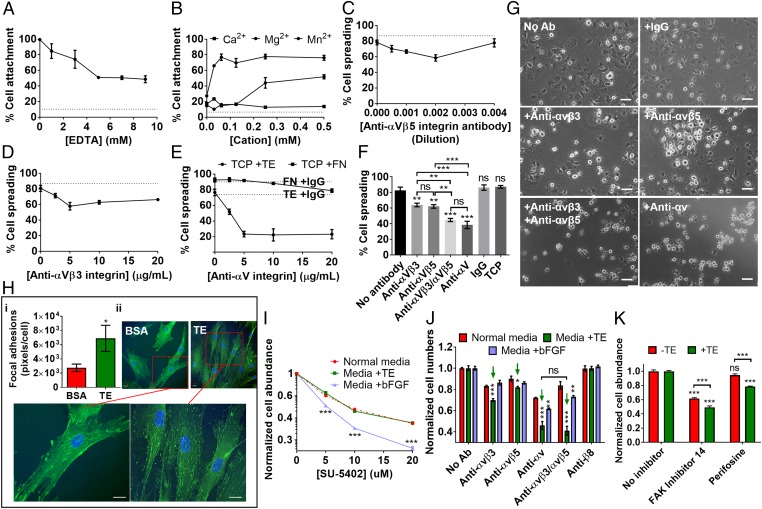
Integrin-mediated effects of tropoelastin on MSC adhesion, spreading, and proliferation. (A) Cell adhesion to substrate-bound tropoelastin in the presence of EDTA. (B) Cell binding to tropoelastin in cation-free buffer with increasing doses of exogenous Mg2+, Ca2+, and Mn2+ divalent cations. (C–E) Cell spreading on tropoelastin with increasing concentrations of an (C) anti-αvβ5, (D) anti-αvβ3, or (E) pan anti-αv integrin antibody. Cell spreading on fibronectin with and without the anti-αv integrin antibody is shown as a control. (F) Cell spreading on tropoelastin in the presence of optimal inhibitory concentrations of anti-αvβ3, anti-αvβ5, combined anti-αvβ3 and anti-αvβ5, and anti-αv integrin antibodies. Cell spreading on TCP and that on tropoelastin in the absence of antibodies or with a nonspecific mouse IgG antibody are also included as controls. Asterisks above the data columns refer to statistical differences from the no-antibody control. (G) Representative images of MSC spreading on tropoelastin, with and without integrin-blocking antibodies. (Scale bar: 100 μm.) (H) Confocal microscope images of MSCs adhered on tropoelastin- or BSA-coated TCP, stained for focal adhesion vinculin (green) and cell nuclei (blue). The relative density of focal adhesion staining per cell is indicated. (Scale bar: 20 μm.) (I and J) MSC proliferation in the presence of (I) FGFR and (J) integrin inhibitors. Cells were grown on TCP in normal media, in media with 20 μg/mL tropoelastin, or in bFGF-supplemented media for 7 d. (I) Increasing doses of the FGFR inhibitor, SU-5402, were added to the media during the proliferation period. Cell numbers were normalized against samples without SU-5402. Cell numbers in media containing tropoelastin or bFGF were compared with those in normal media at each inhibitor concentration to account for the nonspecific toxicity of SU-5402. (J) Optimal inhibitory concentrations of anti-αvβ3, anti-αvβ5, anti-αvβ5 and anti-αvβ3, or anti-αv were added to the media over 7 d. Controls without antibodies or with an antibody against a nonexpressed integrin (anti-β8) were included. Green arrows indicate cells grown in the presence of tropoelastin and αv integrin subunit antibodies. Asterisks above individual columns denote significant differences from cells in normal media at each antibody condition. (K) MSC proliferation after 7 d in the presence of an FAK inhibitor (FAK inhibitor 14) or a PKB/AKT inhibitor (perifosine). Cell numbers were normalized against uninhibited samples. Asterisks above individual columns represent comparison with the no-inhibitor control. *P < 0.05; **P < 0.01; ***P < 0.001; ns, not significant.

