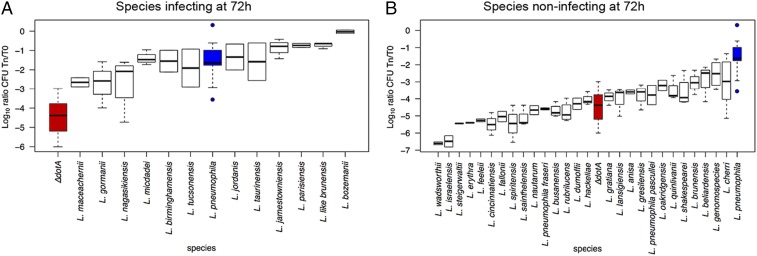
Fig. 5.
The replicative capacity of the different Legionella species in THP-1 cells correlates with their epidemiological features. Replication of each strain at the time point 72 h after infection of THP-1 cells is shown (24 and 48 h postinfection shown in SI Appendix, Fig. S14). Intracellular replication was determined by recording the number of colony-forming units (CFU) after plating on buffered charcoal yeast extract agar. Blue boxes indicate L. pneumophila Paris, representative of a replicating strain; red boxes indicate L. pneumophila ∆dotA, representative of nonreplicating strain. The strains are ordered according to the mean replication values. (A) Legionella species replicating similar to, or significantly better than, L. pneumophila Paris. (B) Species with no replication capacity or significantly lower replication capacity compared to L. pneumophila Paris.

