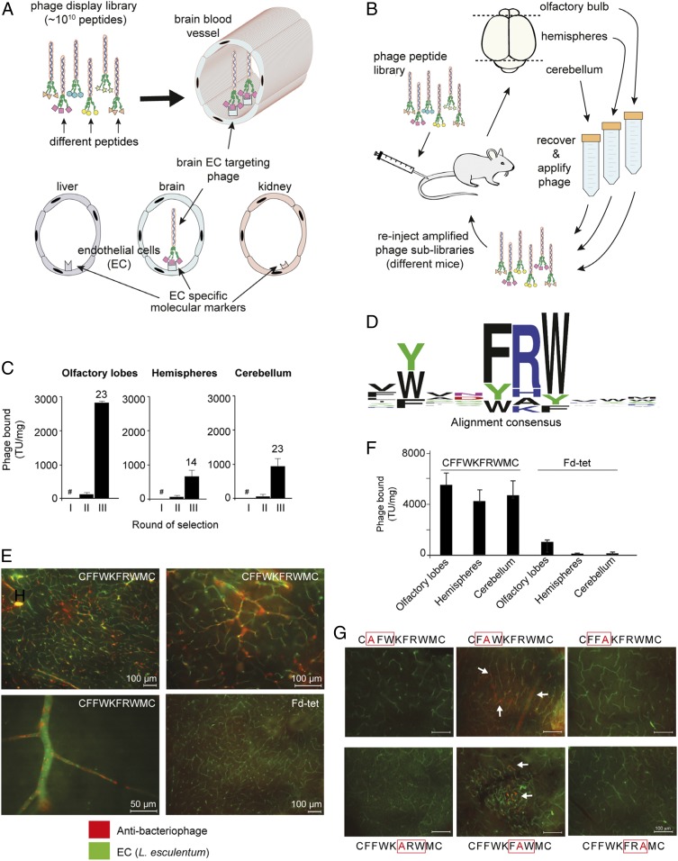
Fig. 1.
Identification of a peptide motif that targets brain blood vessels. (A) Phage display library selection identifies peptides targeting blood vessels. In the CX8C phage library, the ligand peptides are fused to minor capsid protein III (pIII) assembled at one end of the virion. (B) Phage display in vivo scheme. (C) Number of phage particles (TU normalized per milligram of tissue) recovered in each round of selection per brain region. The number above each bar indicates phage enrichment in round III relative to round II; #, round I was not quantified, to minimize loss of unique peptides. (D) Alignment of the total pool of unique peptides (n = 1,021) reveals the consensus motif FRW. (E) Phage CFFWKFRWMC or negative control Fd-tet phage (109 TU) were administered i.v. into mice, and phage bound to blood vessels in brain hemispheres is visualized with antibacteriophage sera (red) and blood vessels with FITC-conjugated Lycopersicon (Tomato) esculentum lectin (green). (Scale bars: 100 µm, unless otherwise indicated.) (F) Quantification by colony count of phage bound to different regions of the mouse brain (n = 4). (G) Effect on phage homing caused by different Ala mutations within the FRW motif. Bars represent means ± SEM from triplicates. (Scale bars: 100 µm.)

