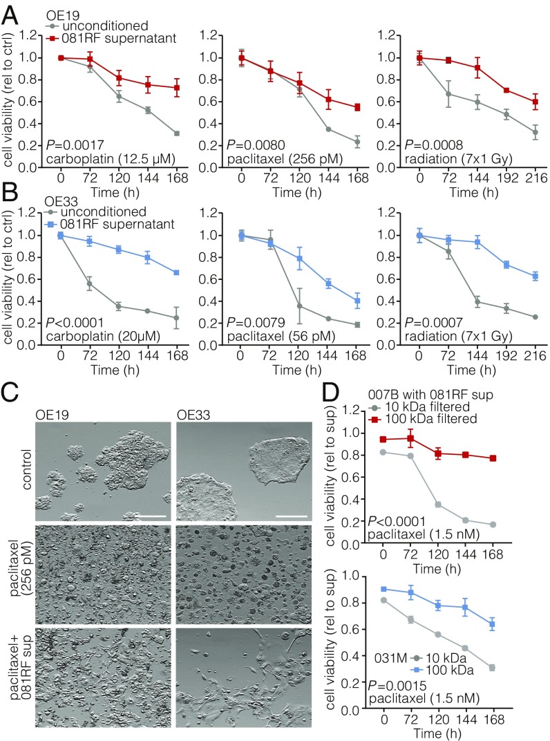
Fig. 1.
Patient-derived EAC-associated fibroblasts confer resistance to chemotherapy and radiotherapy. (A) Cell viability assays were performed and measured at the indicated times, using OE19 cells incubated with the indicated chemotherapeutics and parenthesized concentrations in unconditioned control (ctrl) medium (gray lines) or medium supplemented with 081RF supernatant (1 in 4 diluted) (colored lines). Graphs show means ± SEM of data normalized to t = 0, n = 3. P values were determined by two-way ANOVA and Bonferroni correction. (B) Same as for A, using the OE33 cell line. (C) OE19 and OE33 cell lines were cultured in unconditioned or 081RF supernatant-supplemented medium (081RF sup), treated with 256 pM paclitaxel or control for 168 h, and morphology was assessed by phase-contrast microscopy. (Scale bar: 100 μm.) (D) Cell viability was determined on 007B and 031M cultures which were incubated with 1.5 nM paclitaxel supplemented with 25% 10- or 100-kDa filtered 081RF supernatant. Graphs show means ± SEM, normalized to t = 0, n = 3. P values were determined by two-way ANOVA and Bonferroni correction.

