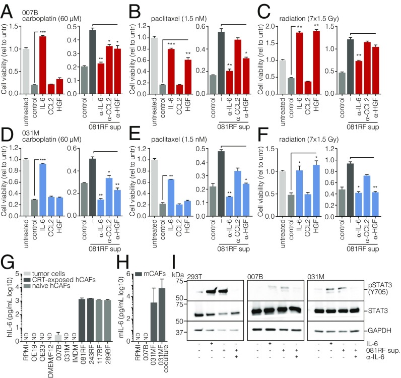
Fig. 2.
Stromal CAF-secreted IL-6 drives therapy resistance. (A–C) Cell viability assays were performed on primary 007B cells incubated for 168 h in the following culture conditions: unconditioned medium without chemotherapeutics (untreated, untr), unconditioned medium with chemotherapeutics (control), conditioned medium with chemotherapeutics, medium supplemented with the indicated cytokines and chemotherapeutics (colored bars), or 081RF supernatant with or without neutralizing antibodies for the indicated cytokines and chemotherapeutics. Graphs show means ± SEM of data normalized to t = 0, n = 3. P values were by one-way ANOVA and compared with the control or 081RF (–) sup only condition. (D–F) As for A–C, using 031M cells. (G) Human IL-6 was measured by ELISA in 3 d-incubated supernatant of the indicated cultures (5 d for 243RF culture) and media not incubated on cells. (H) Mouse IL-6 was measured by ELISA as for G in supernatants from indicated (co)cultures. (I) 293T, 007B, and 031M cells were stimulated for 20 min with medium containing 081RF supernatant incubated for 3 d, diluted 1 in 4. Recombinant IL-6 was used as a positive control, and IL-6–neutralizing antibody was used as a negative control for IL-6–induced STAT3 phosphorylation. Following exposure, cells were lysed and processed for Western blot analysis for the indicated antigens. *P < 0.05, **P < 0.01, and ***P < 0.001.

