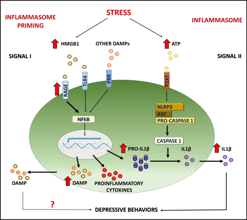
Figure 7. Schematic of stress-induced inflammasome priming.
Exposure to stress activates DAMP-PRR (e.g., HMGB1-RAGE) and downstream NFκΒ signaling to increase transcription of the proinflammatory cytokine IL1β, as well as further increase the expression of HMGB1. Microglial response to stress also requires activation of P2X7 receptors, for example via release of ATP, and activation of the NLRP3 inflammasome complex, which then stimulates the processing of pro-caspase 1 to caspase 1, which in turn cleaves pro-IL1β to IL1β for release. Exposure to CUS results in long-lasting, persistent elevation of HMGB1 and RAGE mRNA expression, and increases sensitivity to subsequent short-term stress exposure, which increases surface/extracellular levels of microglial RAGE. Elevated HMGB1-RAGE underlies increased vulnerability to depressive behaviors such as anhedonia that is blocked in RAGE deletion mutant mice.

