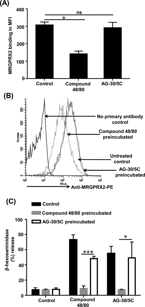
Figure. 5: Compound 48/80 causes MRGPRX2 internalization and reduced MC degranulation whereas AG-30/5C does not.
(A) RBL-MRGPRX2 cells (0.5 × 10⁶) were cultured in the absence (unstimulated) or presence of compound 48/80 (1 μg/mL) or AG-30/5C (1 μM) for 16 h and cells surface expression of MRGPRX2 was determined by flow cytometry using anti-MRGPRX2-PE antibody. The data is presented as mean fluorescent intensity (MFI) of three experiments (B) A representative histogram of cell surface MRGPRX2 receptor expression is shown. (C) LAD2 cells were cultured in the absence (Control) or presence of in compound 48/80 (1 μg/mL) or AG-30/5C (1 μM) for 16h, washed and plated in a 96 well plate (10,000 cells/well). Cells were stimulated with compound 48/80 (1.0 μg/mL) and AG-30/5C (1 μM) respectively for 30 min and percent degranulation was determined by β-hexosaminidase release assay. All the points are expressed as a mean ± SEM of three experiments. Statistical significance was determined by non-parametric t-Test and two way ANOVA. *** indicates P value<.001,** indicates P value <.01 and * indicates P value <.05.

