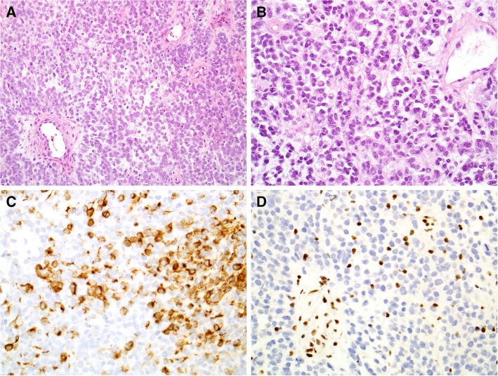
Figure 1.
Histopathology of INI‐1 deficient carcinoma. H&E sections showed sheets of monotonous high‐grade malignant cells (A, ×20) that demonstrated a rhabdoid appearance with eccentric eosinophilic cytoplasm (B, ×40). Immunostains showed positivity for low‐molecular‐weight cytokeratin cocktail CAM5.2 (C, ×40) and loss of integrase interactor 1 expression (D, ×40).

