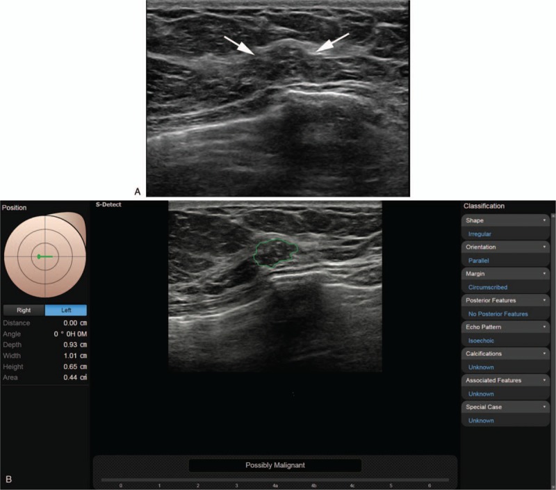
Figure 1.

A. The grayscale ultrasound image in a 57-year-old woman with incidentally detected breast mass on screening examination shows an indistinct irregular heterogeneous hypoechoic mass (arrows) at the 9 o’clock position in the left breast that was diagnosed as breast imaging reporting and data system (BI-RADS) category 3, 4a, and 3, respectively, by less experienced radiologists and 4a and 4b, respectively, by experienced radiologists. B. After review of the CAD application, (where the conclusion was “possibly malignant”), each reviewer recategorized the mass as 4a, 4a, 4b, 4b, and 4c, respectively; core biopsy confirmed the lesion as ductal carcinoma in situ. CAD = computer-aided diagnosis.
