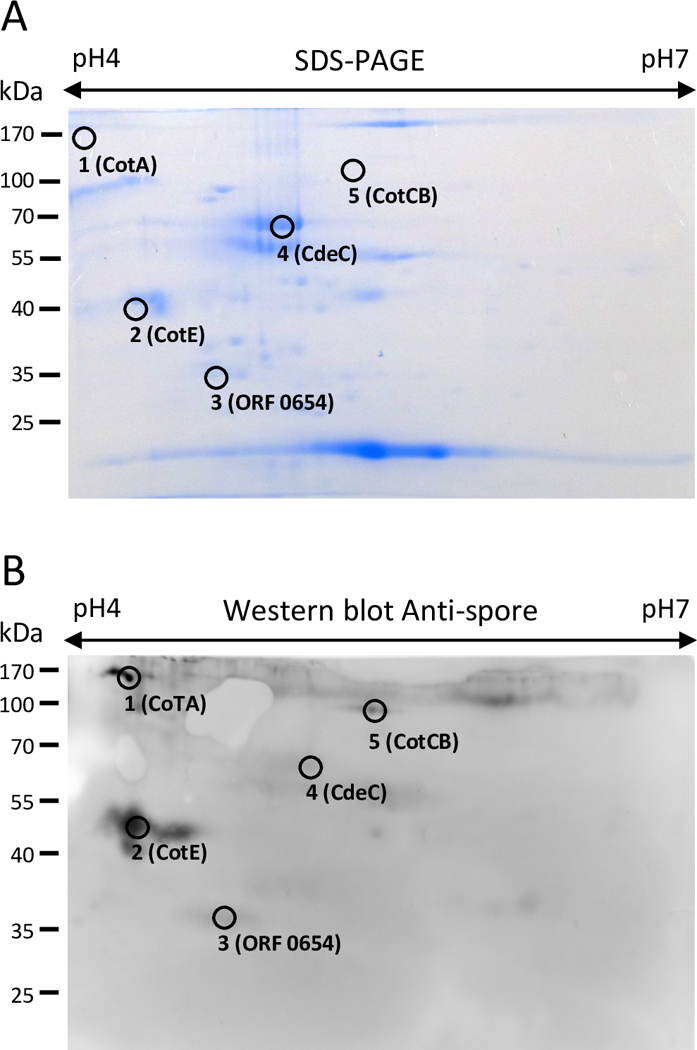
Fig 1. Immunoproteomic analysis of the spore surface of the C. difficile strain R20291 strain.

(A) 2-DE map (pI 4–7) SDS-PAGE of spore coat/exosporium extract with identified immunoreactive proteins stained with Coomassie Brilliant Blue G-250. (B) Western blot of a 2-DE gel of spore coat/exosporium extracts of R20291 epidemic strain probed with anti-spore goat serum specific for C. difficile spores and immunoreactive proteins identified. This gel is representative of three independent 2-DE gels with similar results.
