
Fig. 3.
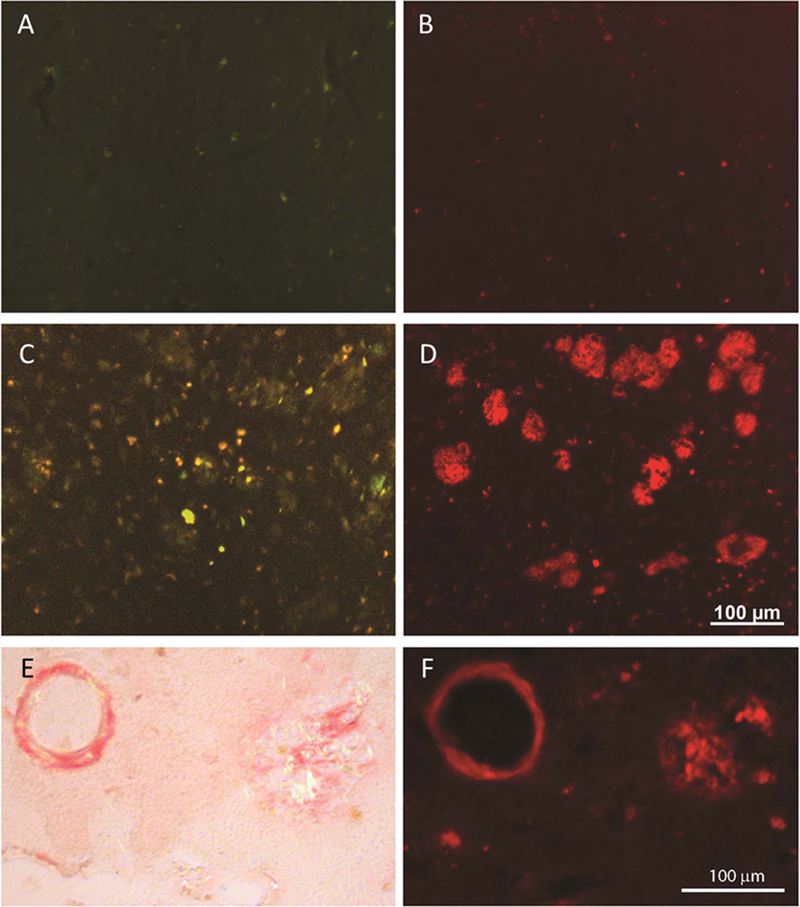
In an aged control individual, the staining with ThS (1.57 mM) (A) and CQ (100 nM) (B) shows very little staining (fluorescence) signal. In AD brain tissue, ThS stains Aβ plaques yet many other fluorescent structures are apparent (C), however, CQ labels the Aβ plaques with greater specificity in the adjacent section from the same case (D). CQ (F) labels the same Aβ plaques and congophilic angiopathy as Congo Red (E).